DermPath Comparison
Recent Submission
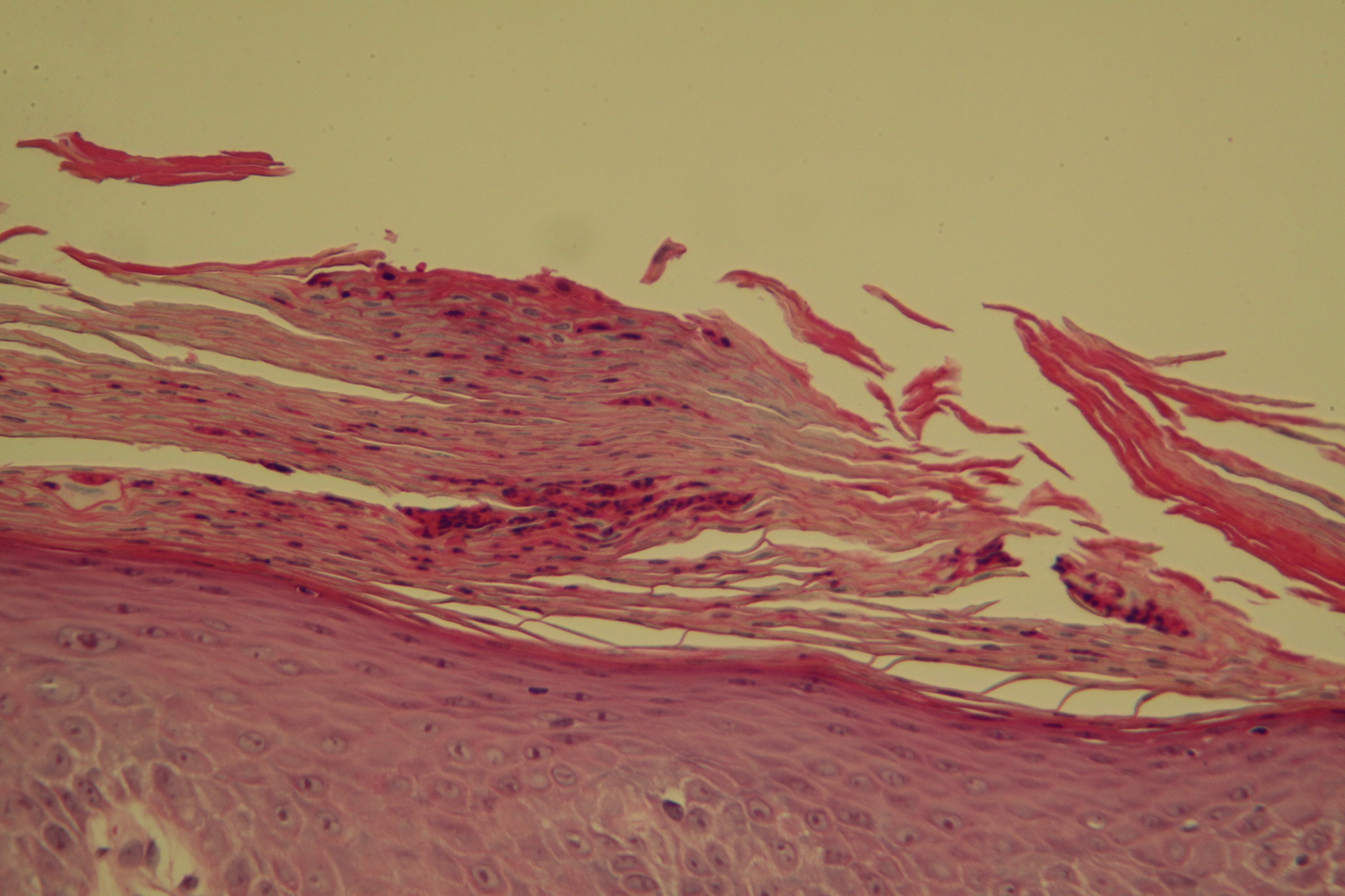
The following table provides a clear comparison of their defining histological features:
| Histological Feature | Psoriasis | Pityriasis Rubra Pilaris (PRP) |
|---|---|---|
| Stratum Corneum (Scale) | Confluent parakeratosis (retained nuclei). | "Checkerboard" pattern: alternating vertical and horizontal bands of orthokeratosis (normal, anucleated cells) and parakeratosis. |
| Granular Layer | Hypogranulosis (thinned or completely absent). | Hypergranulosis (thickened or prominent granular layer). |
| Epidermal Growth | Regular acanthosis: uniform, test-tube-like elongation of rete ridges. | Irregular acanthosis: thickened epidermis with broad, short rete ridges and thick suprapapillary plates. |
| Dermal Papillae | Edematous and elongated. Narrow, elongated dermal papillae. | Broad/thickened rete ridges, narrow dermal papillae. |
| Blood Vessels | Markedly dilated, tortuous, and prominent capillaries. | Mild to moderate vascular dilation. |
| Neutrophils | Often present. Munro’s microabscesses (in stratum corneum) and Kogoj’s spongiform pustules. | Absent or very rare. No migration of neutrophils into the epidermis. |
| Follicles | Follicular involvement is not the primary diagnostic feature. | Prominent follicular plugging (horny plugs filling hair follicles). |
| Dermis | Mixed, sparse to moderate lymphocytic infiltrate. | Sparse, superficial perivascular lymphocytic infiltrate. |
Dermpath Comparison is an online atlas of the Australian Institute of Dermatology. The images may not be used in any other medium without permission from the editors. We have also included here some dermatoscopy comparisons as well.
The Inaugural Editors are:
Dr Ian McColl - Australia
AID DermPath Websites AID Skin Path Atlas AID DermPath Website Ken Paver Project
Dermatoscopy Diagnosis through Structures
Dermoscopy Course of Australian Institute of Dermatology