Psoriasis versus Pityriasis rubra pilaris
Condition 1
Condition 2
Description
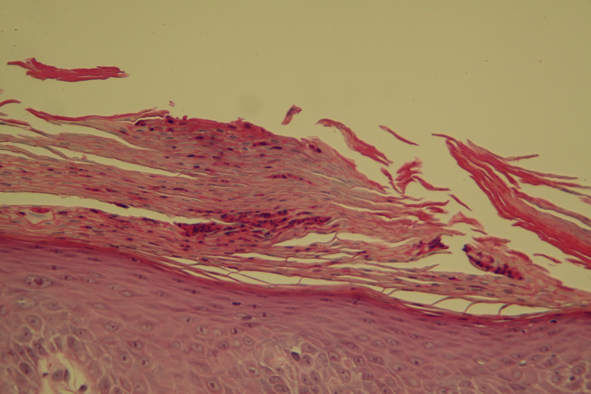
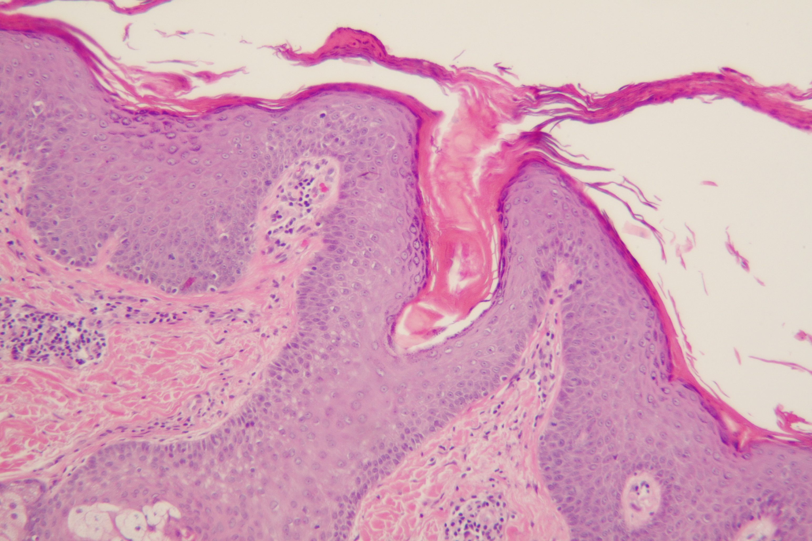
The following table provides a clear comparison of their defining histological features:
| Histological Feature | Psoriasis | Pityriasis Rubra Pilaris (PRP) |
|---|---|---|
| Stratum Corneum (Scale) | Confluent parakeratosis (retained nuclei). | "Checkerboard" pattern: alternating vertical and horizontal bands of orthokeratosis (normal, anucleated cells) and parakeratosis. |
| Granular Layer | Hypogranulosis (thinned or completely absent). | Hypergranulosis (thickened or prominent granular layer). |
| Epidermal Growth | Regular acanthosis: uniform, test-tube-like elongation of rete ridges. | Irregular acanthosis: thickened epidermis with broad, short rete ridges and thick suprapapillary plates. |
| Dermal Papillae | Edematous and elongated. Narrow, elongated dermal papillae. | Broad/thickened rete ridges, narrow dermal papillae. |
| Blood Vessels | Markedly dilated, tortuous, and prominent capillaries. | Mild to moderate vascular dilation. |
| Neutrophils | Often present. Munro’s microabscesses (in stratum corneum) and Kogoj’s spongiform pustules. | Absent or very rare. No migration of neutrophils into the epidermis. |
| Follicles | Follicular involvement is not the primary diagnostic feature. | Prominent follicular plugging (horny plugs filling hair follicles). |
| Dermis | Mixed, sparse to moderate lymphocytic infiltrate. | Sparse, superficial perivascular lymphocytic infiltrate. |
×
![]()