Sweets Syndrome versus Cellulitis
Condition 1
Condition 2
Description
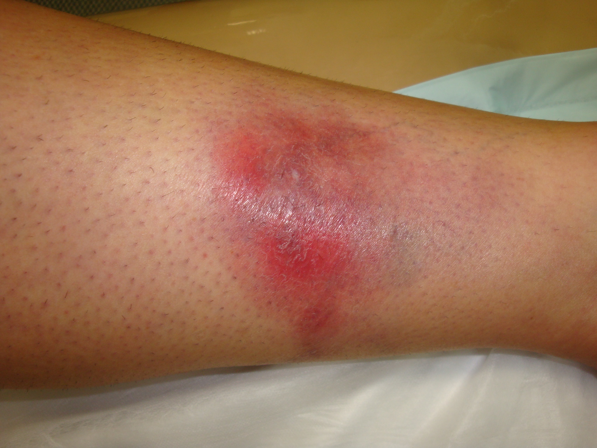
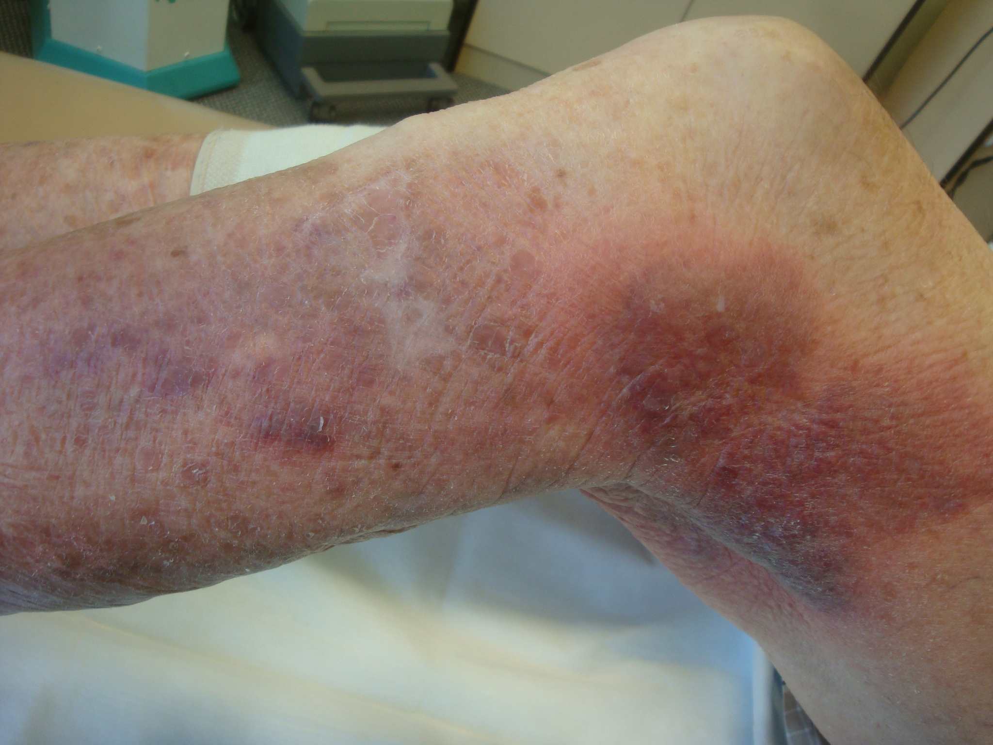
Both conditions can give a tender erythematous plaque on the skin. A history of a predisposing condition such as Crohns or Ulcerative colitis or myeloid disorder helps to point to Sweets but often these patients are on immunosuppressive drugs predisposing them to infections.
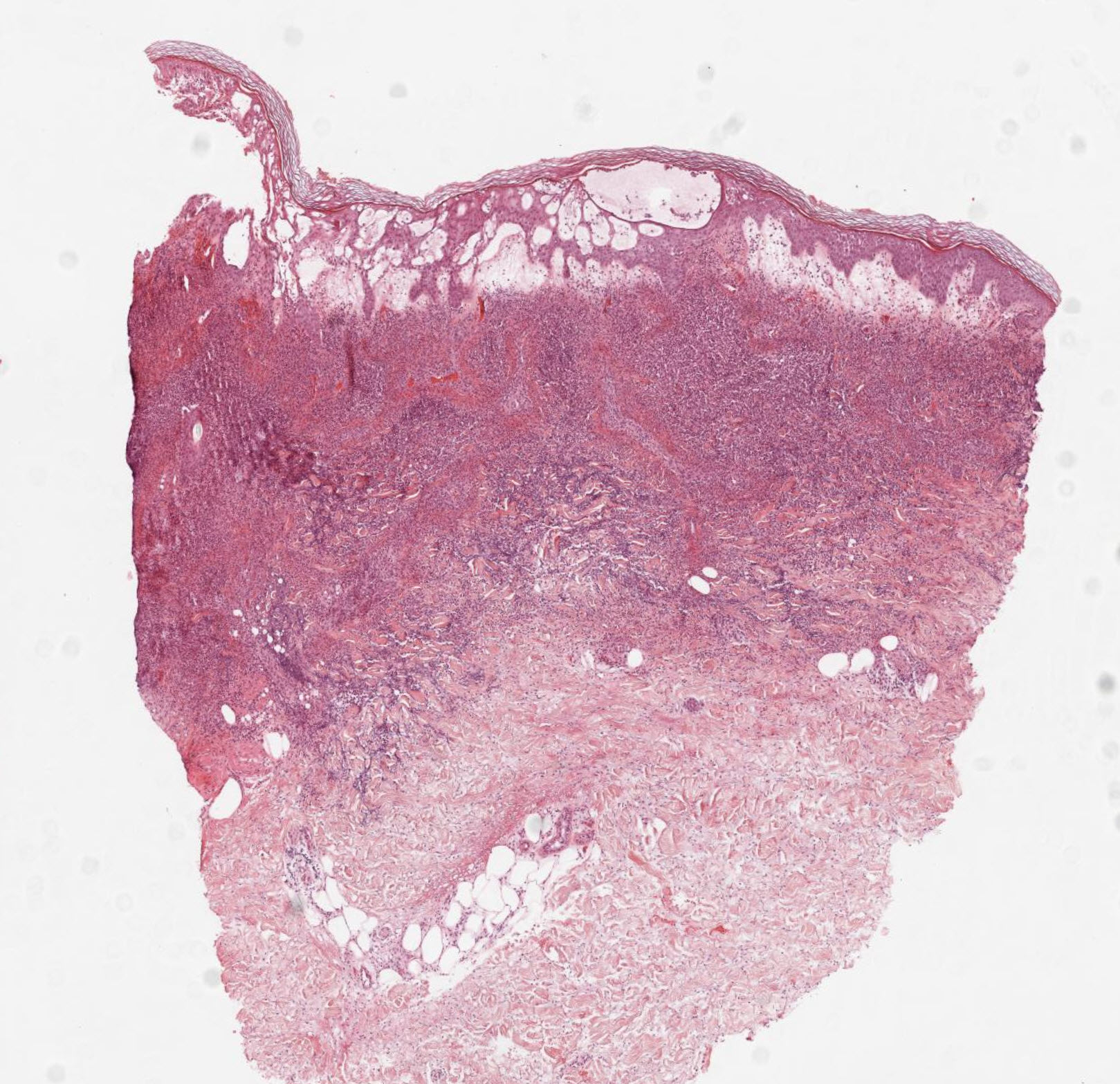
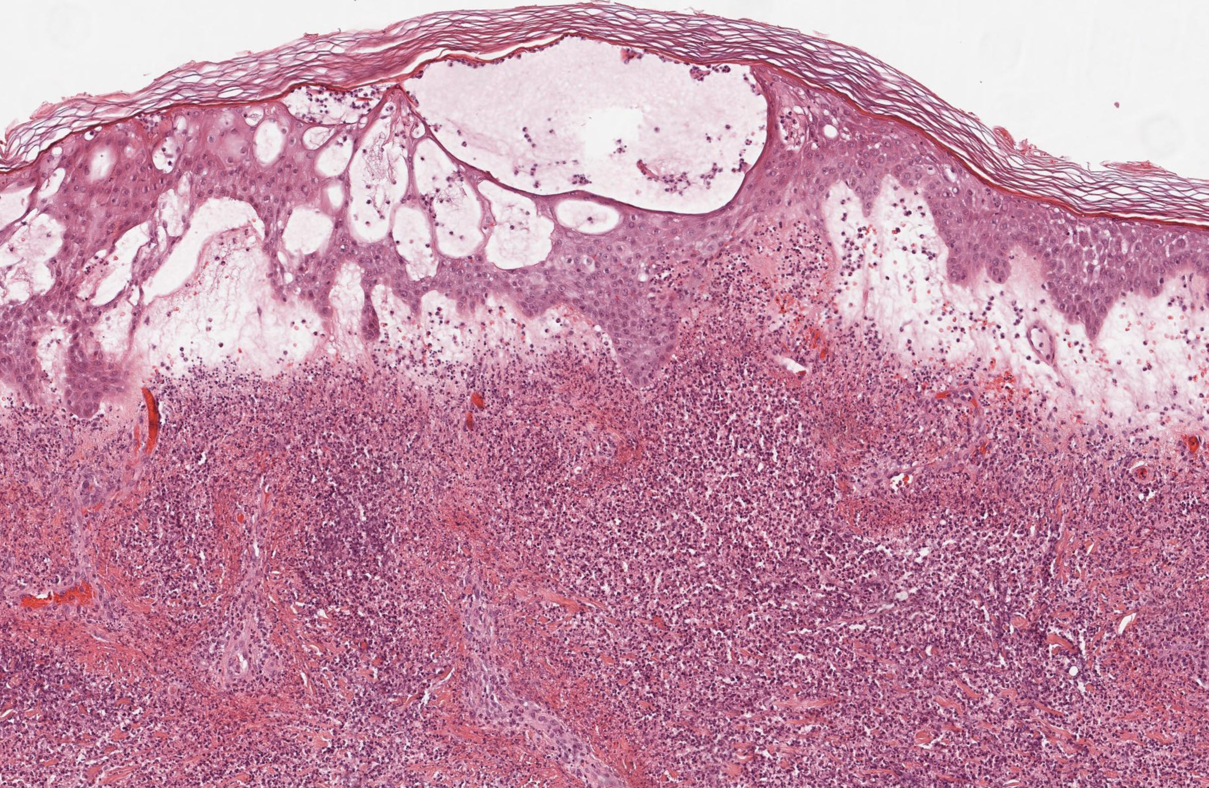
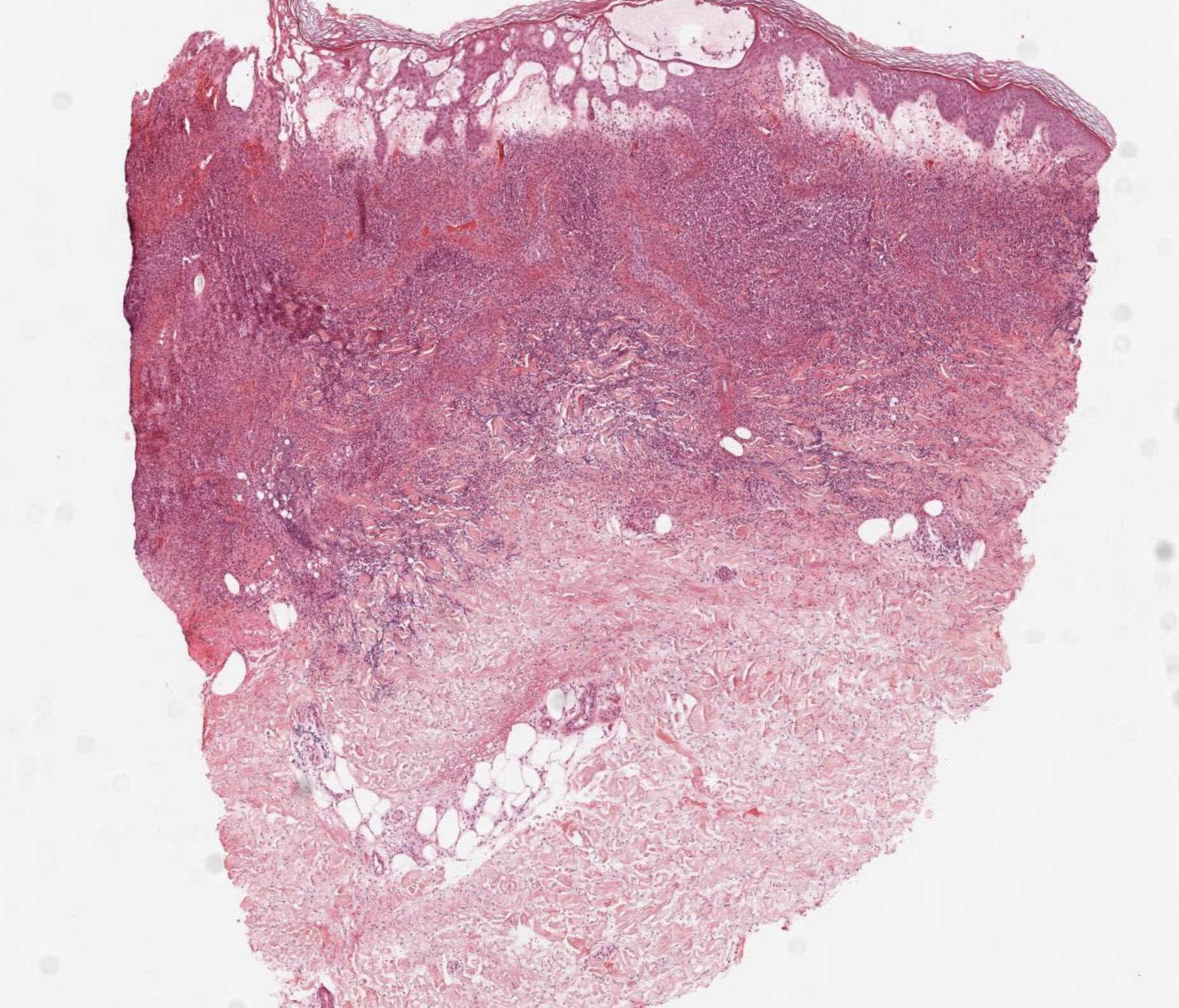
Sweet Syndrome Histology
- Infiltrate Location: Features a dense, diffuse, and nodular infiltrate of mature, neutrophils strictly localized to the upper and mid-dermis.
- Hallmark Feature: Characterized by prominent papillary dermal edema (which gives the lesions a swollen, "juicy" clinical appearance).
- Vasculitis Status: True leukocytoclastic vasculitis (with frank fibrinoid necrosis of the vessel wall) is absent, though neutrophils are often seen clustered around and exiting vessels.
- Micro-abscesses: Subcorneal or intraepidermal pustules may be present.
Cellulitis Histology
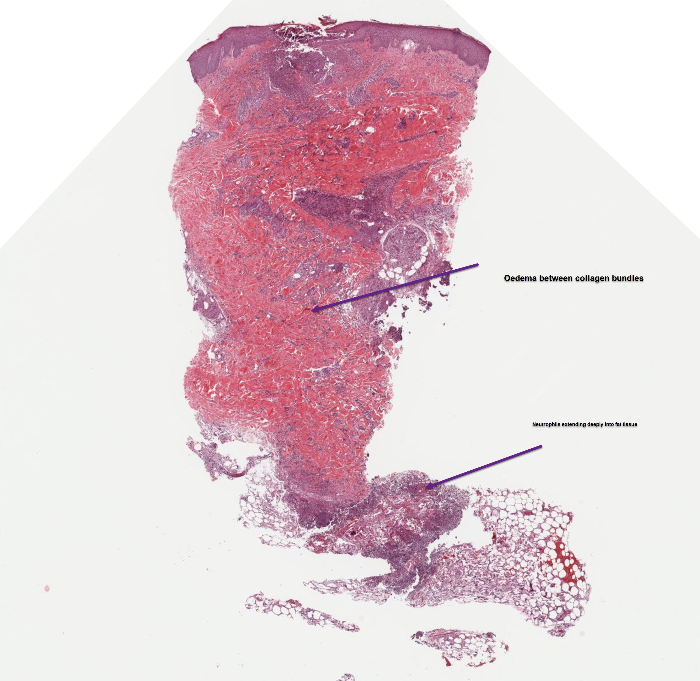
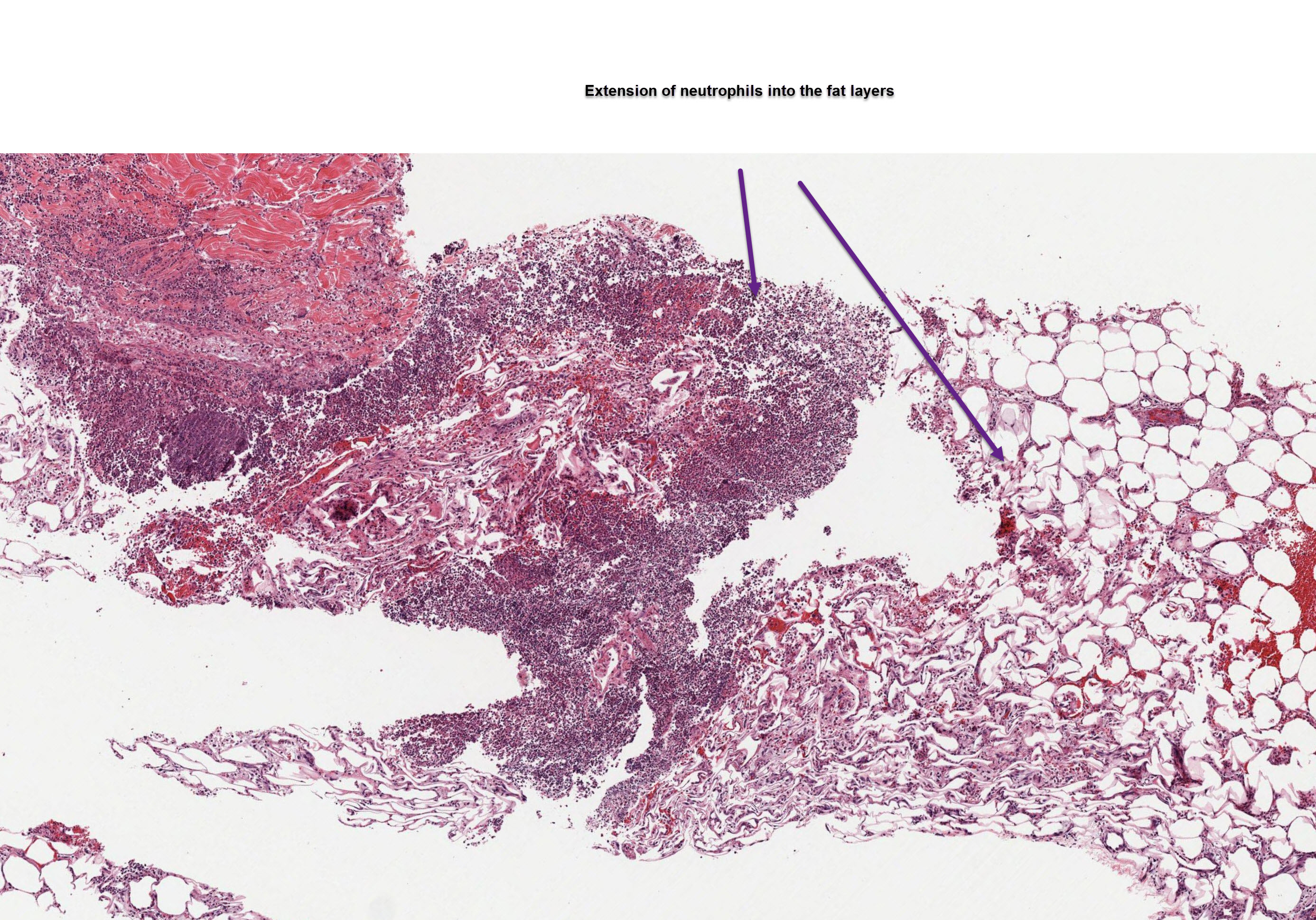
- Infiltrate Location: Features a diffuse, sparse to moderate inflammatory infiltrate that primarily involves the deeper dermis and subcutaneous fat (panniculitis).
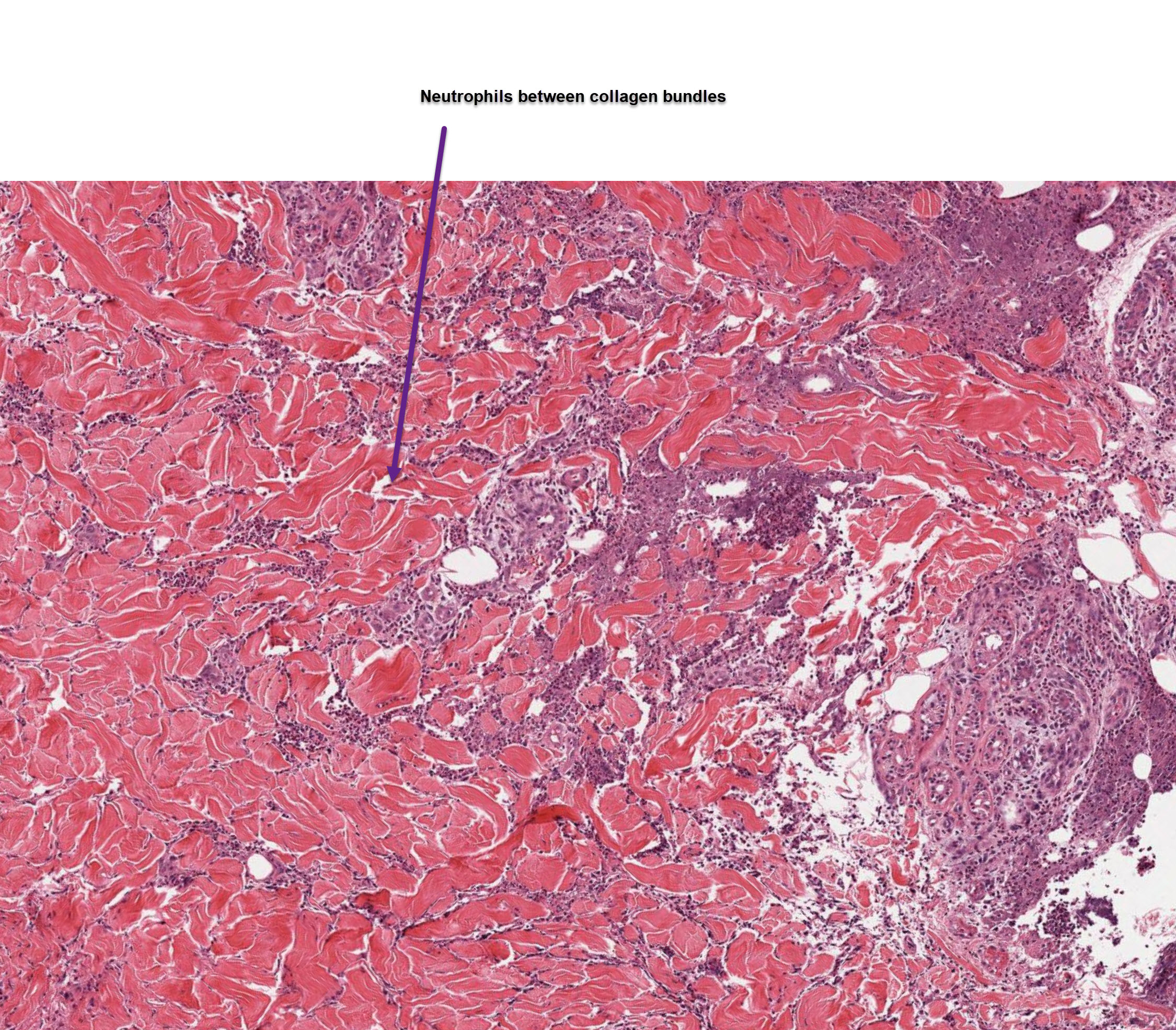
- Cell Types: The infiltrate is often mixed. While neutrophils are present, it characteristically includes lymphocytes, macrophages, and sometimes mast cells.
- Hallmark Feature: Shows signs of acute inflammatory edema spreading diffusely between collagen bundles in the deep dermis.
- Infectious Evidence: Bacterial colonies or structures may be visualized (though often not seen on routine H&E), whereas Sweet syndrome lacks any pathogenic organisms.
Summary of Key Differences
- Depth: Sweet syndrome affects the upper dermis, whereas cellulitis invades the deep dermis and subcutis.
- Edema Type: Sweet syndrome presents with marked papillary dermal edema, while cellulitis presents with widespread diffuse interstitial edema.
- Bacteria: Cellulitis has a bacterial origin (though sometimes subclinical), while Sweet syndrome is an autoinflammatory, sterile neutrophilic dermatosis.
×
![]()